
Bacterial canker of stone fruit, Pseudomonas syringae
Savvas Pastopoulos, Agronomist MSc, Pastopoulos Agronomics, Neos Mylotopos, Pella, Greece
Bacterial canker of stone fruits is caused by bacteria of the genus Pseudomonas and appears as cankers on the trunk, the scaffold limbs and the twigs, with necrosis of the underlying tissue and gum exudation [5]. When the infection girdles a limb, the limb dies back and the leaves turn yellow before necrosis [2], while infected dormant buds are often killed [5]. In Greece the pathovar morsprunorum is recorded in the EPPO database as present and widespread [12].
The plant protection product approvals cited in this article come from the database of the Greek Ministry of Rural Development and Food and are valid in Greece only. Approval of an active substance at Union level does not mean that a given product, crop, target and dose are authorised in any particular country. The national register of the country of use is what applies, and three of the sources below ([16], [17], [18]) are Greek official documents available in Greek only.
Doses of copper are given here per hectare, as the Union legislation states them. The figures measured abroad carry the country and the conditions of measurement inside the sentence, because the timing of the sprays in particular does not transfer from one climate to another.
- The pathogen, two bacteria, not one
- The symptoms
- The annual cycle and the points of entry
- Frost is not a coincidence, the bacterium creates it
- The predisposing factors
- Differential diagnosis, what else looks like this
- Cultivars and rootstocks
- Management
- Copper: which form, when, and why it fails
- When a sample is sent
- Sources
1. The pathogen, two bacteria, not one
The Pacific Northwest plant disease management handbook attributes bacterial canker of cherry to two bacteria and not one: to Pseudomonas syringae pv. syringae and to Pseudomonas amygdali pv. morsprunorum [2].
- pv. morsprunorum, its recorded hosts are Prunus species. The review by Hulin and co-workers gives, for race R1, P. avium, P. cerasus, P. domestica, P. armeniaca, P. persica and P. amygdalus, and for race R2, P. avium, P. cerasus, P. domestica and P. armeniaca [3].
- pv. syringae has a far wider host range. The Washington State fact sheet reports that it attacks sour cherry, peach, plum, small fruits and many weeds found on the orchard floor [1].
Why this matters: because pv. syringae survives on many other plants, the weeds in the rows and the neighbouring vegetation act as an inoculum reservoir [1]. It is no accident that weed control is expressly included among the cultural measures against the disease [2].
In the EPPO database the accepted scientific name remains Pseudomonas syringae pv. morsprunorum, with the code PSDMMP and the authority "(Wormald) Young, Dye & Wilkie"; Pseudomonas mors-prunorum, Pseudomonas morsprunorum and Pseudomonas syringae pv. mors-prunorum are listed as synonyms, while Pseudomonas amygdali pv. morsprunorum does not appear there [11]. There is therefore a divergence between the phytosanitary database and the current phylogenetic literature, and this text states it rather than passing over it.
The address of this article keeps the spelling "morspronorum", because it is already indexed and cannot be changed without breaking links. The correct spelling is morsprunorum, from mors (death) and prunorum (of the plums), and that is the spelling used throughout the text.
2. The symptoms
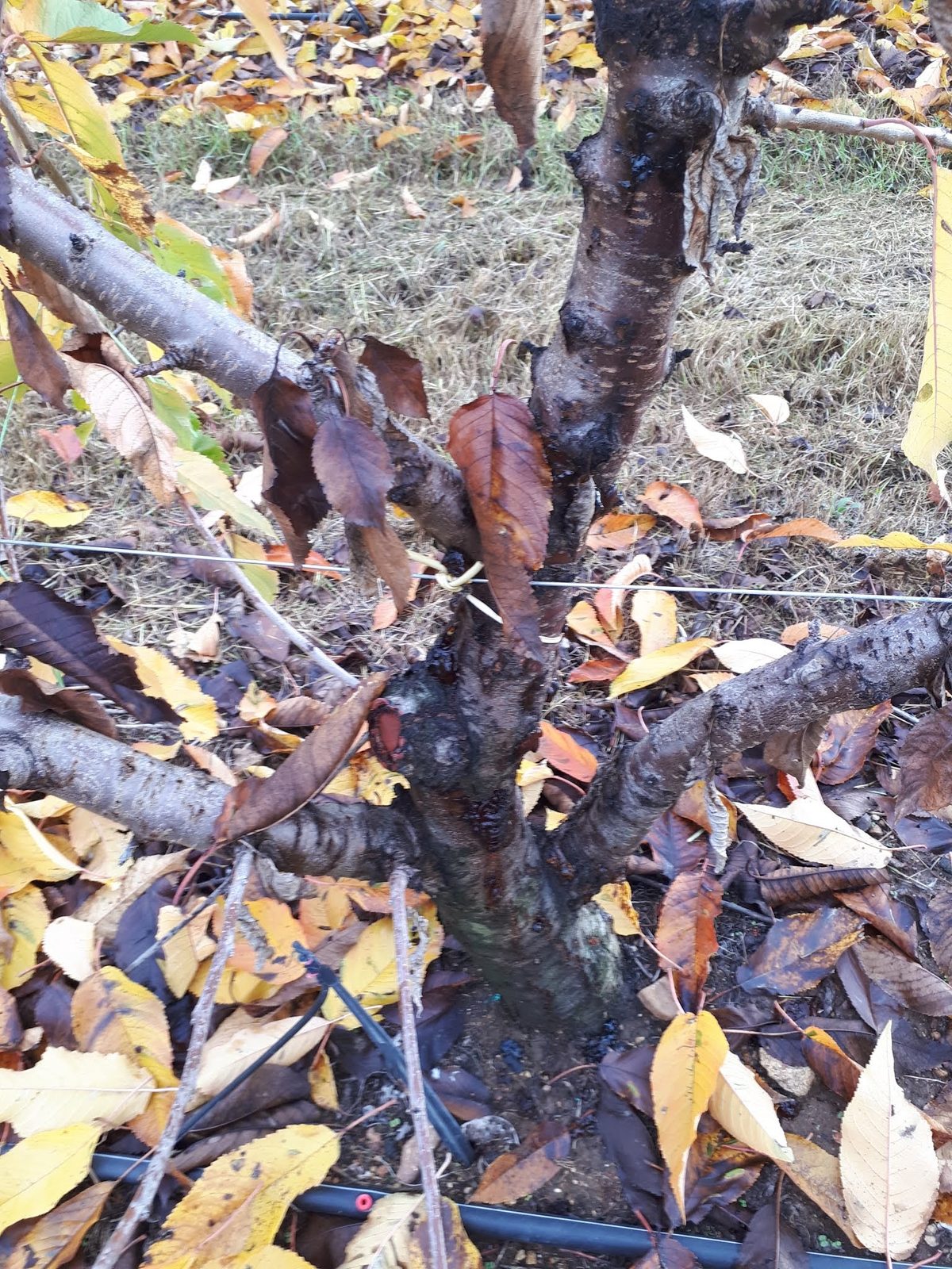
The disease produces a canker that is slightly sunken and darker than the surrounding tissue, on the trunk or on the large scaffold limbs. The inner tissue takes on an orange-brown colour and from the canker narrow brown streaks extend into the healthy tissue, above and below it [5]. The Pacific Northwest handbook describes the affected tissue as discoloured, necrotic cambium [2].
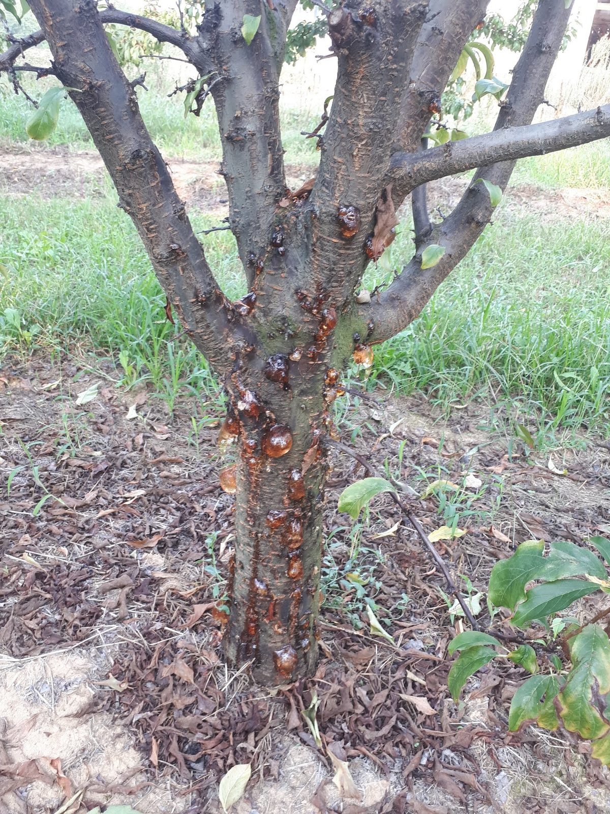
Gum exudation often accompanies the canker, but not always: "a clear to amber exudate, sticky or slimy, may or may not be associated with the cankers" [5].
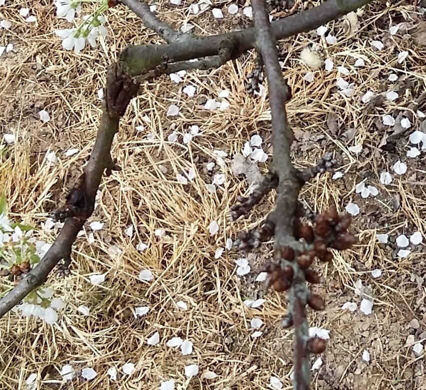
On the buds the picture is distinct and often missed: infected dormant buds are usually killed [5], and the handbook describes "bud necrosis" as a form that starts in spring from the buds of the fruiting spurs and progresses upward in the tree [2].
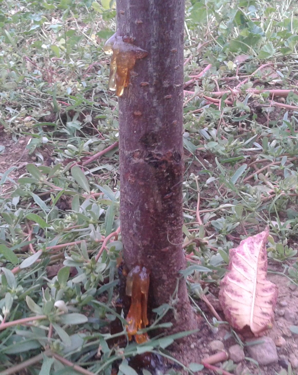
In cherry there are many non-infectious causes of gummosis. The Utah State advisory lists frost cracks, sunburn, bark injury, overwatering, deep planting and severe summer pruning [8]; the Pacific Northwest handbook adds chemical injury, excessive moisture or nitrogen that forces soft growth, and an unsuitable planting site, noting that gummosis "may also be spontaneous" [21].
The colour of the gum does not separate the categories, and the sources themselves do not agree with one another:
- The Utah advisory describes non-infectious gummosis as clear or light coloured, drying to a darker amber over time, and bacterial canker as "milky to clear, light amber gummosis" [8].
- The Oregon publication also gives a clear to amber exudate for bacterial canker [5].
- The Washington State fact sheet, however, describes a dark canker sometimes accompanied by a reddish-brown exudate [1].
- For Cytospora, the 2017 Utah advisory gives dark amber gum [9].
Three sources, three shades for the same pathogen. Colour is an indication, not a criterion.
The reliable criterion in the field is the knife. The instruction is to "use a knife to scrape the outer bark beneath the gum, over a small area" [8]:
- If the inner bark is still cream coloured, the tissue is healthy and the exudate is due to a non-living cause [8].
- If the inner bark is light brown to brown, the tissue is dead and in all probability was killed by a pest or a pathogen [8] [9].
In bacterial canker specifically, the necrosis under the bark shows a distinct reddish-brown pattern affecting the vascular tissue beneath the bark [1], while on the trunk and the scaffold limbs the characteristic feature is the narrow brown streaks extending from the canker into the healthy tissue [5]. One feature that is rarely looked for: reddish lesions with a foul smell on the bark of the root are reported as the picture of root infection by the bacterium [1].
3. The annual cycle and the points of entry
The bacterium overwinters inside the cankers and inside the buds, and these two sites are the main source of inoculum [1]. The population is not constant through the year: it follows a curve with two peaks.
The description in the Washington State fact sheet is explicit: "populations increase in spring after bud break and symptoms develop in spring and summer. By late summer populations decline; however, a second peak of infections may occur in the cooler and wetter conditions of autumn" [1].
The Oregon publication gives the same picture from the detection side: "with the return of autumn rains and lower temperatures, P. syringae is detected at high levels before and during leaf fall" [5].
The optimum temperature range for infection is cool temperatures, 15 to 25 °C (59 to 77 °F in the source), combined with moisture [1]. This is therefore not a disease of hard winter nor of extreme heat, it is a disease of the transitional seasons. Autumn leaf fall, which leaves a multitude of fresh scars exactly when the rains begin, is the most underrated moment of the year [23].
In records from Washington State, USA, infections in new plantings were associated with the heading cut made at planting in spring, when the cut was exposed to irrigation water [1]. The Pacific Northwest handbook gives the corresponding measure in the form of a rule: "heading cuts after planting are made only in dry weather, when no rain is expected for at least a week after pruning" [2].
The remaining documented points of entry in young trees: frost or heat injury on the trunk, wounds from animals and rodents, and the fresh leaf scars, whether from natural fall in autumn or from hand removal of leaves at the base of newly planted trees [1]. The practical conclusion for planting is simple: a heading cut and sprinkler irrigation do not coincide in cold, wet weather, and the trunk of young trees is protected.
4. Frost is not a coincidence, the bacterium creates it
Here lies the most misunderstood mechanism of the disease. The bacterium "can form ice within the plant cell and cause damage, which is called ice nucleation activity (INA)" [1]. The Pacific Northwest handbook puts it from the consequence side: "a protein acts as an ice nucleus, increasing frost injuries, which the bacteria readily colonise" [2].
The quantitative side has been measured: in studies on leaves, "the probability of frost damage, which is proportional to the number of ice nuclei on the leaf surface, is determined to a large extent by the particular bacterial strains" [10]. The same work found that nucleation frequency was generally higher when the strains had grown on the plants than in culture, and that prediction of frost damage must rest on measurements from cells grown on the plant [10].
The cycle closes on itself: the bacterium increases frost injuries, and frost injuries are precisely the entry points through which it infects [2]. The review by Hulin and co-workers confirms it: "environmental factors such as the occurrence of frost can significantly increase the frequency of infection, most probably because of the creation of wounds by frost damage" [3]. That is why choosing a site without frost risk is included among the cultural measures against the disease [2], and why reducing the epiphytic population before the spring frosts has a double benefit.
5. The predisposing factors
The disease rarely strikes on its own. It strikes trees that are already stressed by something else, and the list of cultural measures in the Pacific Northwest handbook is in essence a list of those stresses: choice of a site without frosts, testing the soil for ring nematodes, optimising pH and nutrition, weed control, avoiding interplanting young trees among old ones, delaying winter pruning, and removing affected trees and branches [2].
The ring nematode, recorded in the literature of the period as Macroposthonia xenoplax and today as Mesocriconema xenoplax, was tested on peach trees on two rootstocks, Nemaguard and Lovell, in sandy soil. The result was clear: severe bacterial canker developed only when both organisms were present, and "if either organism was omitted, severe bacterial canker did not develop" [6]. Both rootstocks were good hosts for the nematode and were stunted by it [6].
The mechanism is visible in the root. Ring nematodes "feed mainly on the fine roots, killing them and restricting the development of lateral roots", and that feeding "can lead to the loss of up to 85% of the small roots, causing significant water and nutrient stress" [7].
The practical consequence: the nematode concentrates where the soil texture favours it, "higher populations of Mesocriconema are known to occur in sandy soils with large pores" [7]. On a holding with recurrent bacterial canker, particularly on light, sandy soils, testing the soil for nematodes is part of the programme and not a luxury [2]. Spraying copper alone addresses half the problem. Damage thresholds for the ring nematode are not established even in Washington State [7], and no published Greek threshold was found.
Abiotic factors
- Frosts. Spring frost and severe winter freezes are reported as the main cause of the injury through which infection occurs [5], and infection of cherry increases when the tissue is exposed to extreme temperatures in early and mid winter [5].
- Heat injury on the trunk. It is expressly recorded as a point of entry in young trees [1], while winter injury leading to gummosis is prevented by whitewashing or shading the trunks [21].
- pH and nutrition out of balance. Optimising them is included among the cultural measures against the disease [2].
- Excessive moisture or nitrogen, which force soft growth, and an unsuitable site: cherry trees in wet or unfavourable positions are particularly prone to gummosis [21].
- Overwatering, deep planting and severe summer pruning [8].
6. Differential diagnosis, what else looks like this
The symptoms are not pathognomonic. Gummosis follows other diseases as well, among them brown rot and bacterial canker itself, but it also appears spontaneously [21].
| Pathogen | How it is told apart |
|---|---|
| Bacterial canker P. syringae pv. syringae, P. amygdali pv. morsprunorum |
A canker slightly sunken and darker, with narrow brown streaks extending into the healthy tissue above and below it, and inner tissue that is orange-brown [5]. Under the bark, a distinct reddish-brown necrosis pattern in the vascular tissue [1]. Killed dormant buds [5]. No pycnidia. |
| Cytospora canker Cytospora spp. |
The canker is sunken, with dead tissue at the centre and living tissue at distinct margins that keep extending [19]. On its surface develop scattered raised pimple-like structures the size of a pinhead, the pycnidia, which push out through the bark, and in wet conditions they exude amber to orange, thread-like spore tendrils [19]. This is the most reliable macroscopic distinguishing feature. The foliage wilts and discolours but stays attached, sometimes all winter [19]. Gum may or may not be present [19]. |
| Little cherry and X-disease little cherry viruses, X-disease phytoplasma |
They give small and deformed fruit, poor colour and fruit without flavour [22]. The distribution is the distinguishing feature: "symptoms are confined to one or a few branches, unless the trees have been infected for many years" [22], whereas in bacterial canker the weakened limb is the one that carries the canker and the streaks in the wood [5]. |
| Shothole Wilsonomyces carpophilus |
On the leaves it gives small, round, light brown to purplish spots, rarely larger than 0.6 centimetres (0.25 inch in the source), whose central tissue drops out and leaves the hole [20]. On current-season and one-year-old wood it gives round, reddish, slightly sunken lesions, kills fruit buds and can girdle fruiting twigs [20]. On the fruit the corresponding lesions may produce a clear, sticky exudate [20]. |
7. Cultivars and rootstocks
The differences are of degree, not of absolute immunity. The Washington State fact sheet ranks them as follows:
- Susceptible: Sweetheart, Bing, Staccato, Royal Ann (Napoleon), Van [1].
- Particularly susceptible: Coral Champagne [1].
- With higher resistance: Corum, Regina, Moreau, Lambert, Sam [1]. For Rainier the results are conflicting, with some indication of resistance [1].
The Oregon publication agrees on the substance: "Rainier and Regina appear more resistant than Sweetheart and Bing" [5].
Rootstocks: in Washington State "Mazzard and Colt appear more resistant, while the more dwarfing rootstocks are more susceptible, such as Gisela 6 and Krymsk 5" [1]. The combination of cultivar and rootstock counts for more than either on its own: in Oregon it was recorded that Bing on Gisela 6 suffered 90% mortality and should not be planted in areas where bacterial canker is a problem [5].
And one point that concerns spread within the holding: "vigorous rootstocks have a greater likelihood of natural root grafting and transmission of the pathogen to neighbouring trees than dwarfing and semi-dwarfing ones" [1].
The Greek Ministry's Integrated Crop Protection Guidelines for Sweet Cherry mention, for bacterial canker, the choice of rootstock alone, naming Charger, Mazzard F12-1 and Maxma 14 as relatively resistant [18]. Mazzard is the only rootstock that appears on both sides of the Atlantic as relatively resistant [1] [18].
For the Greek cultivars no published evaluation of susceptibility to bacterial canker was found. The increased susceptibility of Tsolakeika and of Grace Star on wet and heavy ground is recorded here as an observation from practice, without a published Greek measurement, and not as a documented ranking. Likewise, the proportion of the Greek area under each cultivar was not found published.
8. Management
There is no curative chemical control. The Polish research group puts it as a dead end of European practice: "chemical treatments are very limited because of the very small range of bactericides that are available and registered for use in plant production" and "there is no more effective alternative for the control of bacterial diseases" [4]. Control is achieved through preventive measures.
The measures that are documented
- Healthy propagating material and the right site. Choosing a site without frost risk and avoiding the interplanting of young trees among old ones are included among the cultural measures [2].
- Removal of the affected parts. Removal of affected trees and branches and surgical excision of the small cankers in summer [2]; "prune and remove the affected tissues in dry weather, with the cut below the visible canker" [23]. The Greek bulletin calls for complete removal and burning of the affected shoots and branches at pruning [17] and, in the autumn bulletin, removal and burning in dry weather [16].
- Protection of the cuts. The Greek bulletin provides for immediate protection of the large pruning cuts [17] and protection of wounds and cuts with suitable products [16].
- Pruning in dry weather and later in the winter. Delaying winter pruning is an explicit measure [2], and heading cuts after planting are made only when no rain is expected for at least a week [2].
- Testing the soil for ring nematodes before and after planting [2].
- Weed control, since for pv. syringae weeds act as hosts [1] [2].
- Choice of cultivar and rootstock on the basis of documented susceptibility [1] [5].
- Cauterising the cankers. The Washington State fact sheet describes "burning the cankers on the scaffold limbs with a hand-held propane torch", with the note that "it must be done in spring" [1]. It works at the margin of the canker, where the heat reaches and where the woody tissue withstands it.
- Whitewashing the trunk. In new plantings, protecting the trunk with white paint together with a double drip line, so that the water does not touch the trunk, is recorded as good practice [1]. The Pacific Northwest handbook also recommends whitewashing or shading the trunks to prevent the winter injury that leads to gummosis [21]. The measure closes exactly the points of entry: heat injury and cracks from abrupt temperature swings [1].
9. Copper: which form, when, and why it fails
What applies in the European Union
By Commission Implementing Regulation (EU) 2018/1981 the approval of the copper compounds was renewed as candidates for substitution, with an approval date of 1 January 2019. The specific provision sets a maximum of 28 kilograms of copper per hectare over a period of 7 years, while member states may set a maximum annual application rate not exceeding 4 kilograms of copper per hectare. The same regulation requires that the approved quantities should not exceed the minimum necessary to achieve the intended effect [13].
The two figures are consistent with each other and the arithmetic is worth doing once: 28 kilograms spread over seven years averages exactly 4 kilograms per hectare per year, which is the annual ceiling member states may impose. A year at the annual maximum therefore spends a full seventh of the seven-year allowance, and two heavy years in a row leave very little room for the five that follow.
The duration of the approval: the 2018 regulation set an expiry date of 31 December 2025 [13]. The approval has been extended: Commission Implementing Regulation (EU) 2025/1489 of 24 July 2025 replaces, in Part E of the Annex to Regulation 540/2011 and in row 10 "Copper compounds", the expiry date with 30 June 2029 [14]. The limit of 28 kilograms per seven years did not change. The Polish paper of 2023 predicted that copper would leave the list during 2023 [4]; that did not happen, and the example shows why the source for an expiry date must be the implementing regulation itself and not a secondary text.
The active substances that are reported, and what applies here
| Active substance | Union status | What is documented, and what applies in Greece |
|---|---|---|
| Copper hydroxide | A copper compound; approved, candidate for substitution, expiring 30.06.2029 [13] [14] | Recommended as the fixed copper form for the sprays against the disease [1]. In the Polish field trial it was among the most effective products [4]. The Greek bulletins do not name active substances: they refer to "a suitable and approved copper product or a product of another category" [17]. The approval in force, the dose and the pre-harvest interval of each product are checked each time in the approvals database of the Greek Ministry. |
| Copper oxychloride | A copper compound; same status and same limit [13] [14] | In the Polish trial on sour cherry, sweet cherry and plum it was among the products with the highest efficacy [4]. Approval per product and target: the Greek Ministry database [17]. |
| Copper oxide | A copper compound; same status and same limit [13] [14] | The sources disagree. The Washington State fact sheet reports that "copper oxides (Cu⁺) have proved ineffective" [1]. In the Polish field trial, however, copper oxide was among the most effective products [4]. The contradiction is examined below. |
| Bordeaux mixture | A copper compound; same status and same limit [13] [14] | No trial was found that evaluates it separately for bacterial canker of stone fruits. As a copper compound it counts towards the limit of 28 kilograms per hectare over the seven years [13]. For protection of the trunk, the sources consulted report white paint and whitewashing, not Bordeaux mixture [1] [21]. |
| Copper gluconate (foliar fertilisers) | Marketed as fertilisers, not as plant protection products, in the trial consulted [4] | In Poland their efficacy was comparable to that of the copper products: 48.9 to 62.2% on the leaves and 41.7 to 58.3% on the fruit in the 2019 trials [4]. A fertiliser does not replace a plant protection product and carries no plant protection claim; the Greek bulletins refer to approved products [17]. |
| Bacillus amyloliquefaciens QST 713 | Approved in the EU: renewal by Impl. Reg. (EU) 2023/1001, approval from 1.07.2023, expiry 30.06.2038 [15] | In the Polish trial the biological products were clearly less effective than the chemical ones, with variable results for QST 713 [4]. Approval of the active substance in the EU does not imply a Greek approval for this particular target; use presupposes a product approved for the crop and the target [17]. |
| Pantoea agglomerans AM3/18 | A research strain in the trial consulted; no approval act was found | In the Polish trial it showed minimal control of the disease [4]. |
| Fluopyram and fosetyl-Al | A fungicide product tested in Poland against the pathogen [4] | Efficacy 11.9 to 76.9% on the leaves and 36 to 69.3% on sour cherry fruit, similar to or lower than the copper oxide product [4]. No Greek approval for bacterial canker of stone fruits was found; the Greek bulletins refer generally to approved products [17]. |
| Kasugamycin | An antibiotic. The use of the most effective antibiotics is not permitted in the EU [4] | In Washington State it is reported before bloom in combination with copper [1]. Resistance has already been recorded there: "bacteria resistant to Kasumin have been detected in the Pacific Northwest" [2]. It is not an option for the Greek orchard. |
Hydroxide or oxide? The contradiction between the sources
The Washington State fact sheet recommends fixed copper in the hydroxide form (Cu²⁺) and reports that copper oxides (Cu⁺) have proved ineffective [1]. That finding is attributed there to trials by Torres and Latorre on cherry twigs in Chile, published in conference proceedings; the original text could not be opened for the writing of this article, so what is written here comes from the fact sheet's rendering of it [1].
In the opposite direction, the Polish field trial on sour cherry "Nefris", sweet cherry "Van" and plum "Jubileum", with three applications per season before, during and after bloom, found that copper oxide, copper oxychloride and copper hydroxide showed the highest efficacy [4].
The honest formulation: there is a published field trial in Europe that does not confirm the exclusion of the oxide, and a trial on twigs in South America that supports it. The choice of copper form is not a settled question and should not be presented as one. What is common to both sources is that the hydroxide works [1] [4].
Timing, and the absolute limit
- Autumn: in Greece two sprays, at the start of leaf fall and at 75% leaf fall [16]. In the Pacific Northwest the traditional recommendation places the first spray in October, before the autumn rains, with a second application in early January, while an adjustment of the timing to coincide with leaf fall is now proposed [2]. These dates are those of Washington State and Oregon; in Greece the timing is tied to the phenology of the season, as the Greek bulletin specifies [16].
- End of winter: a full cover spray up to bud swell, repeated after 10 to 14 days [17].
- Copper is not applied after full bloom. The instruction is categorical: "do not apply copper products after full bloom" [1]. The reason is not only phytotoxicity: copper harms pollen germination and bees [4].
Why copper fails
Control with copper is unstable in practice. The Pacific Northwest handbook names (1) the copper-resistant strains, "bacteria resistant to copper products have been detected in all cherry growing areas of the Pacific Northwest", (2) poor coverage of the tree by the spray solution and (3) the unfavourable conditions of the season [2]. The same source adds the need to adjust the timing [2].
The uncomfortable record is this: "in small-plot disease control trials, it is not unusual to find trees sprayed with copper with more disease than unsprayed trees" [2]. The Oregon publication says the same: "copper products provide poor control of bacterial canker and in some cases have resulted in more bacterial canker than on unsprayed trees" [5]. And the recent Washington State note: "copper-resistant strains of the bacterium are present, rendering copper sprays ineffective" [23].
There is no Greek datum. No published Greek work was found that tests the sensitivity of Greek strains of the pathogen to copper. The above are American data and they do not transfer silently to the Greek orchard; they do function, however, as a warning about a control programme that in Europe "rests mainly on treatments with copper products" because antibiotics are not permitted [4]. When copper does not deliver, the question is not which product will take its place, but which point of entry was left open.
Sources: 1 2 4 5 13 14 15 16 17 21 23
10. When a sample is sent
Where there is doubt about the pathogen, a sample is sent for laboratory identification. The Benaki Phytopathological Institute accepts diseased plants for phytopathological and entomological examination, together with soil samples for soil analysis and for residues [24]. Outside Greece, the corresponding national diagnostic service applies.
- When the canker has neither the distinct margins and the pycnidia of Cytospora [19] nor the brown streaks extending into the healthy tissue [5].
- When there is general decline with small, poorly coloured fruit and no visible canker, in which case little cherry and the phytoplasma have to be ruled out [22].
- In a new planting with losses of young trees, where the question "did it come with the material or was it infected here?" has financial consequences as well, particularly when planting was accompanied by a heading cut and sprinkler irrigation in cool weather [1].
- When the holding has a recurrent problem despite correctly applied copper sprays, in which case a soil test for nematodes is requested at the same time, this being part of the cultural measures in any case [2] [6].
11. Sources
The numbers in the text refer to the list below. Where only the abstract was used and not the full text, this is stated explicitly.
- [1] Sallato, B., Grove, G. and Johnson, A. Bacterial canker in Washington sweet cherries. WSU Extension Publication FS366E, Washington State University. The main source for the biology and management in Washington State: epiphytic colonisation without symptoms, overwintering in cankers and buds, the requirement for a wound or natural opening together with moisture, the range 15 to 25 °C, the two population peaks, ice nucleation inside the cell, the points of entry in young trees (a heading cut wetted by irrigation, frost, heat injury, animals and rodents, leaf scars), the ranking of cultivars and rootstocks, natural root grafting on vigorous rootstocks, the limited importance of mechanical transmission by tools, the recommendation for copper hydroxide and the report that the oxides are ineffective, the prohibition on copper after full bloom, cauterising with a propane torch in spring, whitewashing the trunk with a double drip line, the host range of pv. syringae and the mention of kasugamycin before bloom. Full text.
- [2] Cherry (Prunus spp.), Bacterial Canker. Pacific Northwest Plant Disease Management Handbook. Source for the two causes of the disease with the name Pseudomonas amygdali pv. morsprunorum, for the protein that acts as an ice nucleus and increases frost injuries, for bud necrosis, for the finding that leaf and fruit spots are uncommon in cherry, for the yellow discolouration before the death of a girdled limb, for the detection of copper-resistant bacteria in all cherry growing areas, for the finding that copper-sprayed trees are sometimes found with more disease than unsprayed ones, for resistance to Kasumin, for the October and early January timing, and for the cultural measures (a site without frosts, testing for ring nematodes, pH and nutrition, weed control, delayed winter pruning, heading cuts only in dry weather with no rain for a week). Full text.
- [3] Hulin, M.T., Jackson, R.W., Harrison, R.J. and Mansfield, J.W. (2020). Cherry picking by pseudomonads: After a century of research on canker, genomics provides insights into the evolution of pathogenicity towards stone fruits. Plant Pathology. Source for the hosts of races R1 and R2 of pv. morsprunorum, for the finding that the two races are distantly related and should not be considered races of one pathovar, for their placement in phylogenetic groups 3 and 1, for the proposal of the name Pseudomonas amygdali for race R1, for the role of the epiphytic leaf population as inoculum for the autumn infections, for the finding that leaf scars are the predominant route of infection for Psm R1 while Pss preferentially invades through wounds, and for the increase in infection frequency with the occurrence of frost. Full text.
- [4] Broniarek-Niemiec, A., Børve, J. and Puławska, J. (2023). Control of Bacterial Canker in Stone Fruit Trees by Chemical and Biological Products. Agronomy 13(4):1166. The only European field trial consulted: studies in central Poland on sour cherry "Nefris", sweet cherry "Van" and plum "Jubileum", against P. syringae pv. syringae and pv. morsprunorum, with three applications per season before, during and after bloom. Source for the finding that copper oxide, oxychloride and hydroxide showed the highest efficacy, for the figures for the copper gluconate fertilisers (48.9 to 62.2% on the leaves, 41.7 to 58.3% on the fruit in 2019), for the figures for the fluopyram and fosetyl-Al mixture, for the clearly lower efficacy of the biological products and the minimal control by Pantoea agglomerans AM3/18, for the statement that the use of the most effective antibiotics is not permitted in the EU and control rests mainly on copper, for the small range of registered bactericides, and for the harm copper does to pollen germination and to bees. Full text.
- [5] Spotts, R.A., Olsen, J., Long, L. and Pscheidt, J.W. (2010). Bacterial canker of sweet cherry in Oregon. Oregon State University Extension Service, EM 9007. Source for the description of the canker as slightly sunken and darker, for the orange-brown inner tissue and the narrow brown streaks extending into healthy tissue, for the clear to amber exudate that may or may not accompany the cankers, for the death of infected dormant buds, for the infection routes (spring frost, severe winter freezes, pruning cuts, insect injury, leaf scars), for the detection of the pathogen at high levels before and during leaf fall, for the increase in infection with extreme temperatures in early and mid winter, for the 90% mortality of Bing on Gisela 6, for the greater resistance of Rainier and Regina compared with Sweetheart and Bing, for the recommendation that cuts be made only in dry weather, and for the finding that copper products give poor control and sometimes more disease than on unsprayed trees. Full text.
- [6] Lownsbery, B.F., English, H., Noel, G.R. and Schick, F.J. (1977). Influence of Nemaguard and Lovell Rootstocks and Macroposthonia xenoplax on Bacterial Canker of Peach. Journal of Nematology 9(3):221 to 224. The study that showed severe bacterial canker developed only with the simultaneous presence of the ring nematode and the bacterium, and that omitting either organism prevented severe disease from developing; rootstocks Nemaguard and Lovell, sandy soil. Full text.
- [7] Plant-parasitic nematodes in pome and stone fruit orchards. WSU Tree Fruit, Washington State University. Source for the feeding behaviour of the ring nematode on the fine roots, for the loss of up to 85% of the small roots with water and nutrient stress, for its co-occurrence with Pseudomonas syringae and cold injury, for the concentration of higher Mesocriconema populations in sandy soils with large pores, and for the finding that damage thresholds are not established. Full text.
- [8] Gummosis of Stone Fruits. IPM Pest Advisories, Utah State University Extension, 4 April 2024. Source for the list of non-infectious causes of gummosis (frost cracks, sunburn, bark injury, overwatering, deep planting, severe summer pruning), for the description of the colour of the gum (clear or light coloured, drying to a darker amber; milky to clear light amber for bacterial canker) and for the knife test: scraping the outer bark beneath the gum, a cream inner bark meaning a non-living cause, light brown to brown meaning dead tissue. Full text.
- [9] Gummosis. IPM Pest Advisories, Utah State University Extension, 18 April 2017. A second, independent statement of the knife test, and source for the dark amber colour of Cytospora gum as against the clear gum of a non-pathogenic cause. Full text.
- [10] O'Brien, R.D. and Lindow, S.E. (1988). Effect of Plant Species and Environmental Conditions on Ice Nucleation Activity of Pseudomonas syringae on Leaves. Applied and Environmental Microbiology 54(9):2281 to 2286. Source for the statement that the probability of frost damage is proportional to the number of ice nuclei on the leaf surface and is determined to a large extent by the particular strains, and for the finding that nucleation frequency is higher in cells grown on the plant than in culture. Full text.
- [11] EPPO Global Database. Pseudomonas syringae pv. morsprunorum (PSDMMP). European and Mediterranean Plant Protection Organization. Source for the name that remains accepted in the phytosanitary database, for the code PSDMMP, for the authority "(Wormald) Young, Dye & Wilkie", for the recorded synonyms and for the finding that Pseudomonas amygdali pv. morsprunorum does not appear there. Full text.
- [12] EPPO Global Database. Pseudomonas syringae pv. morsprunorum, World distribution. Source for the record of the pathogen in Greece with the status "present, widespread", alongside dozens of European countries. Full text.
- [13] Commission Implementing Regulation (EU) 2018/1981 of 13 December 2018 renewing the approval of the active substances "copper compounds" as candidates for substitution (the text of the act as adopted). Source for the maximum of 28 kilograms of copper per hectare over a period of 7 years, for the option for member states to set a maximum annual rate not exceeding 4 kilograms per hectare, for the requirement that the minimum necessary quantities should not be exceeded, for the classification as candidates for substitution and for the dates of approval (1 January 2019) and expiry (31 December 2025). Full text.
- [14] Commission Implementing Regulation (EU) 2025/1489 of 24 July 2025 amending Implementing Regulation (EU) No 540/2011 as regards the extension of the approval periods of certain active substances. EUR-Lex. Source solely for the extension of the approval of the copper compounds: in Part E of the Annex to Regulation 540/2011, row 10, "Copper compounds", the expiry date of the approval is replaced by 30 June 2029. The title, the date and the inclusion of the copper compounds among the substances amended were verified.
- [15] Commission Implementing Regulation (EU) 2023/1001 of 23 May 2023 renewing the approval of the active substance Bacillus amyloliquefaciens strain QST 713. EUR-Lex. Source for the renewal of the approval of strain QST 713, with an approval date of 1 July 2023 and an expiry date of 30 June 2038. Full text.
- [16] ΥΠΑΑΤ, Περιφερειακό Κέντρο Προστασίας Φυτών, Ποιοτικού & Φυτοϋγειονομικού Ελέγχου Βόλου. Γεωργικές Προειδοποιήσεις, Πυρηνόκαρπα Νο 7, November 2024 (Greek Ministry of Rural Development and Food, Regional Centre for Plant Protection, Quality and Phytosanitary Control of Volos, Agricultural Warnings, Stone Fruits No. 7, "Diseases and pests of stone fruits, post-harvest treatments"). The Greek official source for the autumn timing: two sprays, one at the start of leaf fall and one at 75% leaf fall, with the finding that the autumn and winter periods are particularly dangerous, as well as for removal and burning of the affected branches in dry weather and protection of wounds and cuts. The bulletin names no active substances and gives no doses: it refers to products suitable for and approved on the crops. Full text. Available in Greek only.
- [17] ΥΠΑΑΤ, Περιφερειακό Κέντρο Προστασίας Φυτών, Ποιοτικού & Φυτοϋγειονομικού Ελέγχου Βόλου. Γεωργικές Προειδοποιήσεις, Πυρηνόκαρπα Νο 1, 7 February 2025 (the same Greek service, Agricultural Warnings, Stone Fruits No. 1). Source for the end-of-winter timing: a full cover spray with a suitable and approved copper product or a product of another category up to bud swell, repeated after 10 to 14 days; complete removal and burning of the affected shoots and branches at pruning; immediate protection of the large pruning cuts. Here too no active substances are named, which is why every approval, dose and pre-harvest interval is checked in the approvals database of the Greek Ministry. Full text. Available in Greek only.
- [18] ΥΠΑΑΤ. Οδηγίες Ολοκληρωμένης Φυτοπροστασίας στην Κερασιά (Greek Ministry of Rural Development and Food, Integrated Crop Protection Guidelines for Sweet Cherry; finalised December 2013, posted June 2015). Source solely for the Greek recommendation of rootstocks with relative resistance to bacterial canker: Charger, Mazzard F12-1 and Maxma 14. The document gives no active substances, doses or timing for the disease. Full text. Available in Greek only.
- [19] Cherry (Prunus spp.), Cytospora Canker. Pacific Northwest Plant Disease Management Handbook. Source for the features that distinguish Cytospora from bacterial canker: a sunken canker with dead tissue at the centre and living tissue at distinct margins that keep extending, scattered raised pycnidia the size of a pinhead that push out through the bark, amber to orange thread-like spore tendrils in wet conditions, foliage that stays attached, and gum that may or may not be present. Full text.
- [20] Peach (Prunus persica), Shothole. Pacific Northwest Plant Disease Management Handbook. Source for the features of shothole (Wilsonomyces carpophilus): small, round, light brown to purplish spots rarely larger than 0.25 inch, that is about 0.6 centimetres, whose central tissue drops out; round, reddish, slightly sunken lesions on current-season and one-year-old wood, the killing of fruit buds and the girdling of fruiting twigs; a clear, sticky exudate from the fruit lesions. Full text.
- [21] Cherry (Prunus spp.), Gumming. Pacific Northwest Plant Disease Management Handbook. Source for the non-infectious causes of gummosis in cherry (mechanical injury, winter injury, chemical injury, excessive moisture or nitrogen forcing soft growth, an unsuitable planting site), for the finding that gummosis may also be spontaneous while it often follows brown rot and bacterial canker, and for the prevention of winter injury by whitewashing or shading the trunks. Full text.
- [22] Cherry Symptoms. Little Cherry & X-disease Guide, WSU Tree Fruit, Washington State University. Source for the fruit symptoms of little cherry and X-disease (small and deformed fruit, poor colour, lack of flavour) and for the feature that separates them from bacterial canker: the symptoms are confined to one or a few branches, unless the infection has lasted many years. Full text.
- [23] Sallato, B., Serban, C. and Zhao, Y. Effective Strategies for Managing Bacterial Canker During Fall. WSU Tree Fruit, Washington State University (updated 2024, reposted September 2025). Source for the autumn strategy: leaf fall creates a natural wound, and promoting early leaf fall while conditions are dry and warm reduces the risk of infection; avoidance of every injury in wet conditions; pruning and removal of the affected tissues in dry weather, with the cut below the visible canker; and the finding that copper-resistant strains are present and render copper sprays ineffective. Full text.
- [24] Benaki Phytopathological Institute, instructions for the collection and dispatch of samples. Source for the fact that the Institute accepts diseased plants for phytopathological and entomological examination, as well as soil samples for soil analysis and for pesticide residues. Full text. Available in Greek only.
Related articles
Nutrient antagonisms and synergies, with a per-element tool
The arrow diagram in every fertiliser leaflet does not stand up scientifically. The antagonisms are real, but they act on two separate levels, in the soil and inside the plant, and the same element can be antagonistic on one and synergistic on the other. With a tool showing the documented relationships of each element.
Zeolite in agriculture: what is documented and what is not
It is a cation exchanger, it holds ammonium and potassium, not nitrate. The rates that work are tens of tonnes per hectare, not hundreds of kilos. And erionite is a Group 1 carcinogen.
Leonardite in agriculture: properties, rates and what the law says
Leonardite is oxidised lignite with up to 85% humic acids, but pH below 3, up to 6% sulphur and almost no phosphorus. What the analyses and meta-analyses show, where it delivers and where it does not, and what Regulation (EU) 2019/1009 and the organic farming rules require.